
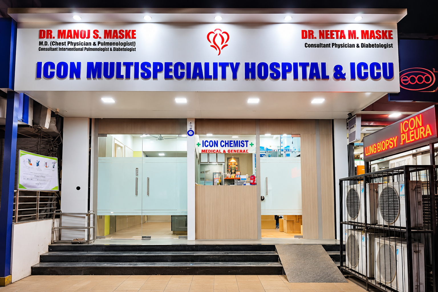
Pleural Diseases Clinic: Ultrasound-Guided Thoracocentesis, Closed Biopsy & Intercostal Drainage
Ultrasound-guided diagnostic thoracocentesis, therapeutic pleural fluid evacuation, and chest tube insertion.
Rapid, ultrasound-guided clinical interventions for fluid accumulation around the lungs (Pleural Effusion), air leaks causing lung collapse (Pneumothorax), pus in the chest cavity (Empyema), and malignant pleural involvement performed with utmost precision by Dr. Manoj S. Maske.
When Is This Care Recommended?
Unexplained Pleural Effusion (Water in Lungs)
Diagnostic sampling to differentiate exudative from transudative fluid and identify the underlying cause.
Spontaneous or Traumatic Pneumothorax
Sudden accumulation of air in the pleural space collapsing the lung, requiring emergent intercostal tube placement.
Empyema (Pus / Infected Pleural Collection)
Thick infected fluid requiring wide-bore or pigtail catheter drainage to prevent fibrothorax and lung entrapment.
Malignant Pleural Effusion with Severe Dyspnea
Rapid therapeutic evacuation of fluid secondary to lung, breast, or metastatic carcinoma providing immediate breathlessness relief.
Tuberculous Pleurisy Evaluation
Analysis of fluid ADA (Adenosine Deaminase), GeneXpert, and pleural biopsy for definitive tubercular confirmation.
Hemothorax (Blood in the Pleural Space)
Immediate evacuated drainage of blood following thoracic trauma or procedural complication to prevent clotting.
Advanced Technology & Clinical Protocols
Real-Time Thoracic Ultrasound Guidance
Visualizing the fluid pocket, diaphragm, and lung parenchyma continuously during needle insertion to eliminate complications.
Pigtail Catheter Seldinger Insertion System
Small-bore, highly flexible drain inserted over a guidewire causing dramatically less discomfort than traditional wide chest tubes.
Standardized Underwater Seal Drainage
Modern, one-way valved drainage systems ensuring continuous unidirectional evacuation of pleural air and fluid.
Comprehensive Fluid Analysis Battery
Immediate testing for protein, LDH, glucose, pH, ADA, GeneXpert, cytology for malignant cells, and bacterial cultures.
Step-by-Step Clinical Journey
Point-of-Care Thoracic Ultrasound Mapping
Scanning the chest wall while the patient sits comfortably to mark the safest, largest fluid pocket with zero organ proximity.
Local Anesthetic Infiltration
Thorough infiltration of skin, intercostal muscles, and parietal pleura with lignocaine, making the procedure virtually painless.
Aspiration or Tube Placement
Gentle aspiration of diagnostic fluid samples or smooth insertion of an intercostal drain connected to an underwater seal bottle.
Post-Procedure Chest X-Ray & Monitoring
Confirming full lung re-expansion and absence of complications before safe transfer or discharge.
Why Choose Icon Lung Clinic for this Speciality?
- Ultrasound-guided technique performed personally by Dr. Manoj S. Maske ensuring maximum safety.
- Substantial reduction in patient discomfort using modern small-bore pigtail catheter sets.
- Rapid diagnostic chemistry and microbiology testing within our on-site pathology laboratory.
- Complete 24x7 emergency capability for tension pneumothorax and massive pleural decompressions.