
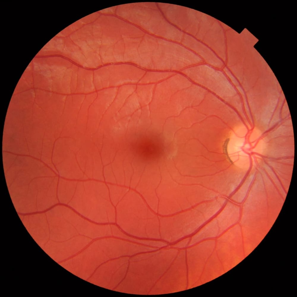
Comprehensive Dilated Retinal Examination & Diabetic Retinopathy Screening
Advanced fundus evaluation to detect diabetic retinopathy, macular degeneration, and retinal microvascular changes early.
Specialized retinal screening and vitreoretinal diagnostics conducted by Dr. Neeta M. Maske. Crucial for detecting diabetic retinopathy, hypertensive retinopathy, age-related macular changes (AMD), and peripheral retinal tears before they jeopardize eyesight.
When Is This Care Recommended?
Diabetes Mellitus (Type 1 or Type 2)
Mandatory annual dilated fundus screening to detect early microaneurysms, hemorrhages, and macular edema.
Systemic Hypertension (High BP)
Evaluation of retinal arteriolar narrowing, nicking, and hypertensive vascular changes reflecting systemic vessel health.
Sudden Floaters, Flashes or Cobwebs
Abrupt emergence of drifting specks, threads, or light sparks indicating vitreous traction or potential retinal tears.
Distorted Straight Lines (Metamorphopsia)
Door frames, window blinds, or straight printed lines appearing wavy or bent, suggesting macular pathology.
Central Dark or Missing Spot (Scotoma)
A blurred or dark blank patch in the center of vision hindering facial recognition and reading.
High Myopia (Power Above -5.00 D)
Elongated eyeballs with thin peripheral retina at elevated risk for lattice degeneration and retinal detachment.
Advanced Technology & Clinical Protocols
Dilated Slit Lamp Biomicroscopy (78D / 90D)
High-magnification stereoscopic examination of the posterior pole, optic nerve head, and foveal reflex using specialized Volk lenses.
Indirect Ophthalmoscopy with 20D Lens
Wide-angle binocular panoramic visualization of the extreme retinal periphery to detect asymptomatic tears and lattice degenerations.
Standardized Amsler Grid Testing
Sensitive bedside tool to detect early central macular swelling, epiretinal membranes, and age-related macular changes.
Collaborative Physician Network
Direct coordination with MD Physician Dr. Manoj Maske for glycemic, lipid, and blood pressure control to halt microvascular leakage.
Step-by-Step Clinical Journey
Pupil Dilation with Gentle Drops
Mild dilating drops administered over 20-30 minutes to expand the pupil for a clear, unobstructed internal view.
High-Magnification Fundus Examination
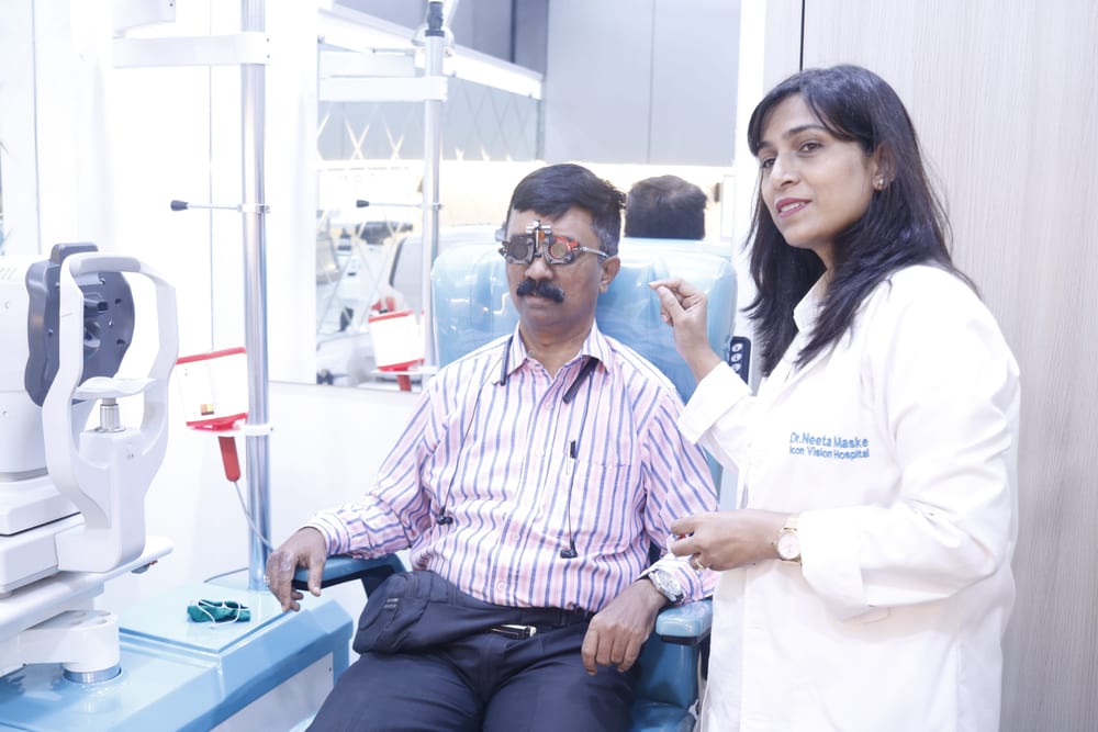
Dr. Neeta Maske inspects the central macula, optic disc, and retinal blood vessels using specialized 78D/90D lenses.
Peripheral Retinal Sweep
Binocular indirect ophthalmoscopy checks the extreme outer retina for holes, lattice degeneration, or tears.
Detailed Report & Treatment Pathway
Clear staging documentation, lifestyle counseling, and referral for retinal laser or anti-VEGF injections if indicated.
Why Choose Icon Vision Clinic for this Speciality?
- Comprehensive diabetic eye evaluations coordinated directly with in-house medical physicians.
- Dilated stereoscopic visualization using imported high-precision optical lenses.
- Early detection of diabetic macular edema preventing permanent central blindness.
- Patient-friendly counseling on blood sugar stabilization, lifestyle, and protective nutrition.